rss
twitter
    Find out what I'm doing, Follow Me :)

Sunday 22 May 2016

INFLAMATION

INFLAMATION


Definition
Inflammation is defined as the local response of living mammalian tissues to injury ie harmful Stimuli such as pathogens or irritant  (Wherein injury is combated and damage repaired). It is body defense reaction in order to elimination the spread of injurious agent as well as to remove the necrosed cells.

Clinical Feature

·         Rubor (Redness)
·         Tumor
·         Color (Heat)
·         Dolor (Pain)
·         Functiolaesa (Loss of Funtion)

Types of Inflammation

Depending upon the defense capacity of host & duration of response, inflammation can be classified as:-
1.      Acute Inflammation.
2.     Chronic Inflammation.

Acute Inflammation

It represent the earliest feature of inflammation response to injury & its duration of response is short, characterized by exudation of fluid & plasma proteins (edema) and emigration of Leukocytes & neutrophils.

Chronic Inflammation   

Inflammation is generally described as chronic wnen it last 6 month or longer.

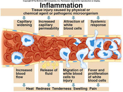
Sequence of event leading to inflamation


  • 1.      Damage to cells, blood vessels and blood components.
  • 2.     Release of vasoactive substances (histamine, serotonin, bradykinin, thromboxanes, prostaglandins)
  • 3.     Vasodilatation of  local capillaries.
  • 4.     Increased permeability of capillaries.
  • 5.     Exudation of fluid into the interstitial spaces.
  • 6.     Fibrin formation through fibrinogen.
  • 7.     Formation of edema/haematoma (causing Tumor or swelling and eventually loss of function)
  • 8.    Fibrinolysis leading to improved circulating.
  • 9.     Proliferation and repair processes.
The changes in acute inflammation can be described under following  two heading:

I) Vascular event  and  II) Cellular event    

I) Vascular event:    

Vasodilation and increased permeability

As defined, acute inflammation is an immunovascular response to an inflammatory stimulus. This means acute inflammation can be broader divided into a vascular phase that occurs first, followed by a cellular phase involving immune cells (more specifically myeloid granulocytes in the acute setting). The vascular component of acute inflammation involves the movement of plasma fluid, containing important proteins such as fibrin and immunoglobulins (antibodies), into inflamed tissue.
Upon contact with PAMPs, tissue macrophages and mastocytes release vasoactive amines such as histamine and serotonin, as well as eicosanoids such as prostaglandin E2 and leukotriene B4 to remodel the local vasculature. Macrophages and endothelial cells release nitric oxide. These mediators vasodilate and permeabilize the blood vessels, which results in the net distribution of blood plasma from the vessel into the tissue space. The increased collection of fluid into the tissue causes it to swell (edema). This exuded tissue fluid contain various antimicrobial mediators from the plasma such as complement, lysozyme, antibodies, which can immediately deal damage to microbes, and opsonise the microbes in preparation for the cellular phase. If the inflammatory stimulus is a lacerating wound, exuded platelets, coagulants, plasmin and kinins can clot the wounded area and provide haemostasis in the first instance. These clotting mediators also provide a structural staging framework at the inflammatory tissue site in the form of a fibrin lattice - as would construction scaffolding at a construction site - for the purpose of aiding phagocytic debridement and wound repair later on. Some of the exuded tissue fluid is also funneled by lymphatics to the regional lymph nodes, flushing bacteria along to start the recognition and attack phase of the adaptive immune system.
Acute inflammation is characterized by marked vascular changes, including vasodilation, increased permeability and increased blood flow, which are induced by the actions of various inflammatory mediators. Vasodilation occurs first at the arteriole level, progressing to the capillary level, and brings about a net increase in the amount of blood present, causing the redness and heat of inflammation. Increased permeability of the vessels results in the movement of plasma into the tissues, with resultant stasis due to the increase in the concentration of the cells within blood - a condition characterized by enlarged vessels packed with cells. Stasis allows leukocytes to marginate (move) along the endothelium, a process critical to their recruitment into the tissues. Normal flowing blood prevents this, as the shearing force along the periphery of the vessels moves cells in the blood into the middle of the vessel.
II) Cellular event
The cellular component involves leukocytes, which normally reside in blood and must move into the inflamed tissue viaextravasation to aid in inflammation. Some act as phagocytes, ingesting bacteria, viruses, and cellular debris. Others release enzymatic granules that damage pathogenic invaders. Leukocytes also release inflammatory mediators that develop and maintain the inflammatory response. In general, acute inflammation is mediated by granulocytes, whereas chronic inflammation is mediated by mononuclear cells such as monocytes and lymphocytes.

a)Leukocyte extravasation

Various leukocytes, particularly neutrophils, are critically involved in the initiation and maintenance of inflammation. These cells must be able to move to the site of injury from their usual location in the blood, therefore mechanisms exist to recruit and direct leukocytes to the appropriate place. The process of leukocyte movement from the blood to the tissues through the blood vessels is known as extravasation, and can be broadly divided up into a number of steps:

1) Leukocyte margination and endothelial adhesion: The white blood cells within the vessels which are generally centrally located move peripherally towards the walls of the vessels. Activated macrophages in the tissue release cytokines such as IL-1 and TNFα, which bind to their respective G protein-coupled receptors on the endothelial wall. Signal transduction induces the immediate expression of P-selectin on endothelial cell surfaces. This receptor binds weakly to carbohydrate ligands on the surface of leukocytes and causes them to "roll" along the endothelial surface as bonds are made and broken. Cytokines from injured cells induce the expression of E-selectin on endothelial cells, which functions similarly to P-selectin. Cytokines also induce the expression of integrin ligands such as ICAM-1 and VCAM-1 on endothelial cells, which mediate the adhesion and further slow leukocytes down. These weakly bound leukocytes are free to detach if not activated by chemokines produced in injured tissue. Activation increases the affinity of bound integrin receptors for ICAM-1 and VCAM-1 on the endothelial cell surface, firmly binding the leukocytes to the endothelium.

2) Migration across the endothelium, known as transmigration, via the process of diapedesis: Chemokine gradients stimulate the adhered leukocytes to move between adjacent endothelial cells. The endothelial cells retract and the leukocytes pass through the basement membrane into the surrounding tissue using adhesion molecules such as ICAM-1.

3)Movement of leukocytes within the tissue via chemotaxis: Leukocytes reaching the tissue interstitium bind toextracellular matrix proteins via expressed integrins and CD44 to prevent them from leaving the site. A variety of molecules behave as chemoattractants, for example, C3a or C5, and cause the leukocytes to move along a chemotactic gradient towards the source of inflammation.

b)Phagocytosis

Extravasated neutrophils in the cellular phase come into contact with microbes at the inflamed tissue. Phagocytes express cell-surface endocytic pattern recognition receptors (PRRs) that have affinity and efficacy against non-specific microbe-associated molecular patterns (PAMPs). Most PAMPs that bind to endocytic PRRs and initiate phagocytosis are cell wall components, including complex carbohydrates such as mannans and β-glucans, lipopolysaccharides (LPS),peptidoglycans, and surface proteins. Endocytic PRRs on phagocytes reflect these molecular patterns, with C-type lectin receptors binding to mannans and β-glucans, and scavenger receptors binding to LPS.
Upon endocytic PRR binding, actin-myosin cytoskeletal rearrangement adjacent to the plasma membrane occurs in a way that endocytoses the plasma membrane containing the PRR-PAMP complex, and the microbe. Phosphatidylinositol and Vps34-Vps15-Beclin1 signalling pathways have been implicated to traffic the endocytosed phagosome to intracellular lysosomes, where fusion of the phagosome and the lysosome produces a phagolysosome. The reactive oxygen species,superoxides and hypochlorite bleach within the phagolysosomes then kill microbes inside the phagocyte.

Phagocytic efficacy can be enhanced by opsonization. Plasma derived complement C3b and antibodies that exude into the inflamed tissue during the vascular phase bind to and coat the microbial antigens. As well as endocytic PRRs, phagocytes also express opsonin receptors Fc receptor and complement receptor 1 (CR1), which bind to antibodies and C3b, respectively. The co-stimulation of endocytic PRR and opsonin receptor increases the efficacy of the phagocytic process, enhancing the lysosomal elimination of the infective agent.
Saturday 14 May 2016

Pain-Inflamation

Pain-Inflamation


It is an unpleasant stimulus, a very common sensation experienced by each one of us. Pain is our body’s way of sending warning to our brain. The degree to which one feels pain and one’s reaction to it is extremely variable, depending on our biological, psychological and cultural makeup. Each one of us has a different threshold for pain. Awareness of pain is a complex experience of sensing, feeling and thinking.

Pain occurs in many different types such as sharp, jabbing, throbbing, burning, stinging, tingling, nagging, dull and aching. Pain also varies in intensity, varying from mild or acute to severe or chronic. Severe pain is profoundly disturbing and generally produces a greater physical and emotional response than mild pain. Severe pain can also be incapacitating and interfere with our daily routine, compelling the patient to seek immediate relief usually from a doctor.

Nerve ending beneath our skin sense heat, cold, touch, pressure and pain. Whenever there is an injury to our body, these nerve endings get stimulated and will send impulses to our spinal cord and to our brain. Pain involves three major components of our nervous system :1)Peripheral nerves, 2) Spinal cord & 3) Brain.

1)   Peripheral Nerve :

They form a network of nerve fibers that spreads throughout body. At the ends of some of these special nerve endings “nocciceptors”, that can sence an unpleasant stimulus, such as a cut, burn or pressure. Nociceptors are most crowded in our skin, bones, joints, muscles and in the protective membrane around our internal organs. They can also sence pressure, temperature, chemical changes, detect inflammation caused by injury, disease or infection.

2) Spinal Cord:

When pain messages reach the spinal cord, they come across specialized nerve cells that act as “gatekeepers” and filter the massages that reach the brain. For severe pain that can cause body harm, e.g. touching a hot stove or when a pin pricks, the “gate” is opened wide and the massages delivered rapidly to our brain. Nerve cells in the spinal cord also respond to these urgent warnings. These cells in spinal cord trigger some other cells of the nervous system, such as motor nerves (reflex action). Our motor nerves cause our muscles to pull our hand away from the burner or pain. Weaker pain messages are filtered or blocked out at the gate.

3) Brain :

When pain messages climb up from spinal cord to reach the brain, they arrive at the thalamus, a sorting and switching station. The thalamus quickly interprets the messages as pain and forwards the impulses simultaneously to three specialized regions of our brain:
1. Physical sensation region (somatosensory cortex)
2. Emotional feeling region (Limbic system) and
3. Thinking region (Frontal cortex)
Our brain responds to the pain message by promoting healing eg. If we cut our finger, brain signals the autonomic nervous system to send additional blood and nutrients to the site of injury.

Natural Pain killers and pain enhancers

Painkillers:

Our brain and spinal cord produce their own painkillers called Endorphins or enkephalins, similar to morphine, a narcotic drug often used to treat severe pain. When released, these Chemical attach to spinal receptors in our brain, producing “stop-pain” messages.

Pain enhancers:


A protein called substance P stimulates nerve ending i.e nocciceptors at the site of injury to increase pain messages. Other pain enhancers work by activating normally silent nerve cells in the injured area thereby prompting these cells to amplify pain messages.

Saturday 7 May 2016

ASHWAGANDHA (as a powerful herb in Ayurvedic Medicine)

ASHWAGANDHA
 A powerful herb in Ayurvedic Medicine. 


It Also known commonly as  Indian ginseng, poison gooseberry, or winter cherry. Aswagandha, one of the most powerful herbs in Ayurvedic healing, has been used since ancient times for a health benefits. Sanskrit Name: -Ashwagandha- Means 'the smell of a horse' as its fresh root smells like a horse's urine, and also perhaps because it is renowned for imparting the sexual stamina of a horse.


Synonyms:                                                

                  Withania Root, Asgandh

Biological Source:

It consists of dried roots and stem bases of “ Withania somnifera” belonging to family Solanaceae.

Geographical Source:

This plant grow wildly in all drier part and subtropical India. It occurs in Madhya Pradesh, Uttar Pradesh, Punjab and north western parts of India like Gujrat and Rajasthan. It is also found in Congo, South Africa, Egypt, Morocco, Jordan, Pakisthan and Afganistan.

Chemical Constituents:

The Mail constituents of ashwagandha are alkaloids and steroidal lactones.
Main Alkaloid – Withanine.

Other Alkaloids – Somniferine, Somnine, Somniferinine, Withananine, Pseudo-withanine, Tropine, Pseudo-Tropine, Choline, Isopelletierine and Anaferine.

The leaves contain steroidal lactones, which are commonly called as
“withanolides”.

Uses :

  • Ashwagandha is effective for insomnia but does not act as a sedative and hypnotic effects.
  •  Its rejuvenative and nervine properties produce energy which in turn help the body to settle and sleep. A herb that rejuvenates the nervous system, erases insomnia and eases stress.
  •  It has Hypotensive Respiratory stimulant actions along with dradycardia.
  •  It is an immuno – modulatory agent.
  •  Ashwagandha has also been shown to lower blood pressure and is highly effective in stopping the formation of stress induced ulcers.
  •  Improves learning, memory, and reaction time
  •  Reduces anxiety and depression without causing drowsiness
  •  Helps reduce brain-cell degeneration
  •  Stabilizes blood sugar
  •  Helps lower cholesterol
  •  Offers anti-inflammatory benefits
  •  Contains anti-malarial properties
  •  Enhances sexual potency for both men and women-
Ayurvedic uses

Traditionally, it has been used in treatment of Rheumatism, Gout, Hypertension, Nervine, and Skin disease. It has been widely used as sex-stimulant and Rejuvenator and is considered as strength and vigour promoting drug especially in geriatric cases.

Ashwagandha Preparations : -
Ashwagandha is used in Ayurvedic medicine in many different ways. Some of the more common methods include as a Ashwagandha powder/ Ashwagandha churna, Ashwagandha extract , a decoction, medicated ghee -Ashwagandha ghruta, as a medicated wine- Ashwagandharishta, Ashwagandha rasayana, Ashwagandhadi churna.


Ayurvedic properties of Ashwagandha:

Vajikara- Increases sexual desire 
Rasayani- Rejuvenates the body 
Balya- Increases strength 
Ati shukrala- Improves quality and quantity of semen 
Shwitrapaha- Useful in management of white discoloration of the skin 
Shothahara- Useful in management of edematous conditions.it helps clear impurities (Ama) from the various channels of the body. 
Kshayapaha- Useful in treating emaciation and under nutritive conditions 

Bio energetics:
Rasa (Taste)- Tikta (Bitter); Katu (Pungent); Madhura (Sweet) 
Guna (Characteristics)- Laghu (Light); Snigdha (Unctuous) 
Veerya (Potency)- Ushna (Warm) 
Vipaka (Post digestion effect)- Madhura (Sweet)
Effect on Tridoshas (Three bio humors): 
Pacifies Kapha and Vata Doshas i.e. it is useful in management of all diseases originating from aggravated Kapha and Vata.


Ashwagandha as an Adaptogenic Herb

In Ayurveda, there are specific herbs with adaptogenic benefits, Ashwagandha is one of them. Adaptogens are substances (a combination of amino acids, vitamins, and herbs) that modulate your response to stress or a changing environment. Adaptogens help the body cope with external stresses such as toxins in the environment and internal stresses such as anxiety and insomnia.

Sunday 1 May 2016

Guduchi as a natural drug


Guduchi (A crud drug)
A valuable Remedy in Ayurveda

Synonyms :

Giloe, Gulancha 


Guduchi (Tinospora cordifolia), also known as amrit, is one of the most valued herbs in the Ayurvedic pharmacy. According to myth, when the ancient gods churned the primordial ocean, an ambrosial nectar was created that would grant immortality to any who drank it. The nectar was named amrit, a Sanskrit word that means “imperishable.

Biological Source:


Leave & dried stem of plant “ Tinospora Cardifolia”.

Family : Menispermacceae

Chemical Constituents:

  • Diterpenoids – Tinosporaside, Tinosporide, Cardifolioside A & B, Coloumbin
  • It also contains – Jatrorrhizine, Berberine, Palmitine, Protoberberine

Uses :

  • Anti-inflammatory, Analgesic and Antipyretic.
  • Reduction in fasting blood sugar.
  • Used in Diarrhea and Dysentery.
  • Anti-stress activity and useful in anxiety and illness.
  • Decrease blood pressure.
  • Decrease blood urea level.
  • Preventing cold and flu.
  • Immune system enhancement.
  • Hepatitis and Jaundice (Protect the liver from exposure to toxins).
  • Chronic skin disorders such as psoriasis or eczema.
  • Reducing the side effect of chemotherapy drugs.

Guduchi and the Doshas

Amrit contains the bitter, pungent, and astringent tastes. Although it’s traditionally used to remove accumulated Pitta, guduchi can balance all the doshas.
Combined with other herbs:
Guduchi can be combined with shatavari or ashwagandha as a general tonic, or with aloe vera juice for the purposes of detoxification. –
  • Guduchi can be combined with shatavari or ashwagandha as ageneral tonic.
  • Guduchi with aloevera juice can be used as detoxification.
  • Guduchi along with aloevera and papaya leaves are very useful in dengue fever.


Monday 25 April 2016

LINEZOLID - A ANTIBIOTICS OF OXAZOLIDINONE GROUP

LINEZOLID
 A ANTIBIOTICS OF OXAZOLIDINONE GROUP


Linezolid a antibacterial agent of  a new class of antibiotics, know as Oxazolidinone used for treatment of Gram-positive bacterial infection that are resistant to other antibiotics.

Linezolid is active against most Gram-positive bacteria that cause disease, including Streptococci, Vancomycin-resistant Enterococci(VRI), methicillin-resistant staphylococcus aureus (MRSA), Streptococcus pneumoniae (penicillin-susceptible strains only), Staphylococcus epidermidis(including methicillin-resistant strains), streptocovvus pyogenes, & Enterococcus faecalis. The main uses are infections of the skin and pneumonia.

Mechanism of Action:

Linezolid are a protein synthesis inhibitor that it stops the growth of bacteria by disrupting their production of proteins. Although many antibiotics work this way but linezolid appears to be unique in that it blocks the initiation of protein synthesis and not one of the later steps; therefore, cross-resistance between linezolid and other classes of antibiotics is unlikely.

Pharmacokinetics:

Plasma concentrations of linezolid at steady-state after oral doses of  600 mg given every 12 hours(q12 h).

Absorption :

Linozolid is rapidly and extensively adsorbed after oral dosing. Maximum plasma concentrations are reached approximately 1 to 2 hours after dosing and the absolute bioavailability is approximately 100%. Therefore, linezolid may be given orally or intravenously without dose adjustment. It may be administered without regard to the timing of meals. The time to reach the maximum concentration is delayed from 1.5 to 2.2 hours and Cmax is decreased by about 17% when high fat food is given with linezolid.

Distribution:

Linezolid readily distributes to well-perfuse tissue. The plasma protein binding of it is approximately 31% and is concentration-independent.

Metabolism :
Linezolid in not detectably metabolized by human cytochrome P450 and it does not inhibit the activities of clinically significant human CYP isoforms .

Excretion:

Nonrenal clearance accounts for approximately 65% of the total clearance of linezolid. Under steady-state conditions, approximately 30% of the dose appears in the urin as linezolid, 50% as metabolite. The renal clearance of linezolid is low (average 40 ml/min) and suggests net reabsorption. Virtually no linezolid appears in the feces.

Dosage:

Adult- 600 mg orally every 12 hours.

Geriatric:

The  pharmacokinetics of linezolid are not significantly altered in elderly patients (65 yrs or older). Therefore, dose adjustment for geriatric patients is not necessary.

Pediatric:

Information indicates that pediatric patients dosed with 10mg/kg IV have a similar Cmax but a higher average clearance when corrected by body weight and shorter apparent elimination half-life than adults receiving 625 mg of linezolide. Studies with dose higher than 10mg/kg or more frequent than every 12 hours have not been conducted in pediatric patients.

Gender:

Females have a slightly lower volume of distribution of linezolide than male. Plasma concentrations are higher in female than in males ,which is partly due to body weight difference. After a 600mg dose, mean oral clearance is approximately 38% lower in females than in males. However, there are no significant gender difference in means apparent elimination –rate constant or half-life. Therefore, dose adjustment by gender does not appear to be necessary.

Renal Insufficiency:

Linozolide pharmacokinetics is not altered in patients with any degree of renal insufficiency; no dose adjustment is recommended for patients with renal insufficiency.

Hepatic Insufficiency:

The pharmacokinetics of linezolide in not altered in patients with mild-to-moderate hepatic insufficiency. On the basis of the available information, no dosage adjustment is recommended for patients with mild-to-moderate hepatic insufficiency.

Pregnancy: Teratogenic Effects:


Linezolide was not teratogenic in mice or rate at exposure levels 4-fold (in mice) the expected human exposure level.However, embryo and fetal toxicities were seen there are no adequate and well-controlled studies in pregnant women. Linezolide should be used during pregnancy only if the potential benefit justifies the potential risk to the fetus

Sunday 17 April 2016

Nosocomial Infections

Nosocomial Infections


Infections which are acquired from hospitals are called nosocomial infection. These infection occurring with in 48 hours of hospital admission, 3 day of discharge or 30 days of an operation. If the organisms come from another patient it is called cross infections and if patient himself carries the infection to some other site then it is autoinfection. Infection may become apparent during the stay of the patient in the hospital or after his discharge from the hospital. There is actual increase in frequency and severity of infection especially due to antibiotic resistant enterobacteria, Staphylococcus aureus and Pseudomonas aeruginosa.Thus prolonged stay of the patient in the hospital is undesired and may be a serious matter for the patient and his family.

Patients Requiring Isolation:

Some patients really need isolation. Patients of tuberculosis, typhoid, diphtheria, lassa fever or smallpox should not be treated or nursed in open ward as these disease are serious and easily transmissible. Similarly infants with measles or whooping cough should not be nursed in general ward but may be treated at home. Staphylococcus aureus infection cases especially resistant to many antibiotics belonging to phase types (80/81 or 75/77) capable of causing serious epidemic of hospital sepsis, certainly require isolation. Isolation cubicles are suggested for these purposes which should be so designed, equipped and managed that no microorganism can pass from them to a ward. Attendant should use gown on entering the cubicle and remove on leaving. Washing facilities for the patient and attendant of the patient must be provided in the cubicle. Dressing should be discarded into paper bags which may be removed to incinerator. Bedding and clothing should be kept in disinfectant solution before sending to laundry. When the patient finally leaves the cubicle,it must be thoroughly washed with disinfectant and all equipment must be sterilized as far as possible.

Hospital Infection and Prevention:

We should be aware of some important hospital infections and their prevention:

  1. Wounds and burns: It is important to remove all tissue debris from accidental wounds and burns as bacteria can establish more easily in damaged tissue. A careful and aseptic technique for dressing of wound preferable in dressing room reduces chances of cross infection.
  2. Urinary tract infection: Catheter or other instruments into the bladder may cause urinary tract infection. Used catheters are difficult to sterilize and may be the cause of cross infection also, hence disposable sterilized catheter should be used aseptically.
  3. Alimentary tract infections: Outbreak of E.coli gastroenteritis in children and Shigella sonnei, dysentery do occur quite oftenly  in hospital. Isolation, general hygiene and exclusion of carriers are important preventive measures.

Epidemiological Markers Useful in investigating Hospital Infection:

    • Antibiogram and resistogram.
    • Biotyping.
    • Phage typing.
    • Bacteriocin typing.
    • Serotyping.
    • Serum opacity factor.
    • RNA electropharesis as in done in rotavirus.
    • Cytotoxicity assay, e.g. Proteus mirabilis.
    • Plasmid profile.

Prevention of Nosocomial Infections:

  • Proper washing
  • Isolation of patients, e.g. plague, influenza, measles, etc.
  • Careful and appropriate use of instruments.
  • Use of antibiotics only if required. It may be given to carrier staff or patient.
  • Use of blood transfusion only if must. Disinfectants of excreta and infected material.
  • Surveillance of infection properly and regularly.
  • Use of vaccine, e.g. tetany gas sangrene, hepatitis – B, etc.

Factors Responsible for Hospital Infections:

  • Neonates and aged patients have risk of getting hospital infection because of long stay and decreased immunity.
  • Impaired defense mechanisms of patients due to disease or treatment.
  • Hospital environment contains relatively heavy load of microorganisms.
  • Major invasive diagnostic or therapy procedures.
  • Advance treatment of cancer, organ transplantation, etc.
  • Presence of multidrug resistant bacteria, etc.

Source of Hospital Infection :

  • Infecting microorganisms from fellow patients which may be multidrug resistant.
  • Infected organisms from hospital staff
  • Infecting organisms from instruments, blood products, intravenous fluid, etc.
  • From patient’s normal flora, etc.
  • Insects are also source multidrug infection.
  • Organism may be present in air, dust, water, antiseptic solution, food, etc.
  • Surface contaminated by patient’s secretions, blood fluid, etc.

Mode of Infection :


  • Airborne.
  • Contact, e.g. hand, clothing, etc.
  • Food and water.
  • Hospital equipments and instruments.
  • By parenteral routs.